
Gradual visual change in adulthood can result from glaucoma, but it can also come from cataract, an incorrect spectacle prescription, diabetic eye disease, retinal disease or another optic-nerve condition. You will learn which symptom patterns need same-day care, what an evaluation measures, and how doctors decide whether drops, laser or surgery can protect the vision you still have.
Key takeaways
- Arrange an eye examination for gradual side-vision loss, even without pain.
- Visual fields and OCT can detect glaucoma damage before you notice symptoms.
- Ask doctors to check for cataract, retinal disease and refractive error too.
- Treatment protects remaining vision; it cannot restore optic nerve damage already lost.
Does gradual vision change fit glaucoma, or does it need emergency care?
Glaucoma often damages peripheral vision before central vision, so you can read and recognise faces while losing side vision unnoticed. That damage can progress without pain. Blur, glare, reduced contrast and difficulty seeing in dim light point to other possibilities, including cataract or refractive error, but gradual change still needs an eye examination.
- Book an examination for gradual change, even when you have no pain. Glaucoma treatment for gradual vision changes in Mumbai cannot be chosen from symptoms alone.
- Seek same-day emergency eye care for sudden vision loss or sudden loss of part of your field of view.
- Treat a painful red eye, severe headache with nausea or vomiting, or halos around lights as an emergency. Acute angle closure is possible, and a routine appointment is not the right level of care.
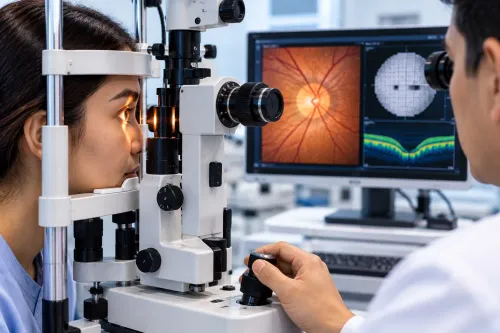
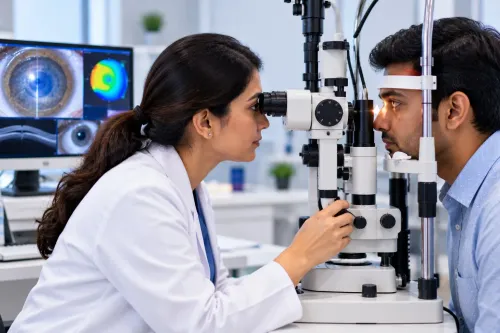
An assessment combines visual acuity, slit-lamp examination, eye-pressure measurement, optic-nerve evaluation, visual-field testing and gonioscopy; OCT and corneal-thickness measurement often add useful evidence. One normal pressure reading does not rule out glaucoma. The specialist must identify open-angle, angle-closure or secondary disease, including causes linked to steroid exposure, trauma, inflammation or lens problems.
What happens during an assessment for adult glaucoma?
The appointment starts with medical and family history, including diabetes, steroid exposure, eye trauma, inflammation, lens-related problems and a close relative with glaucoma. Tell the clinician about tablets, inhaled steroids, creams near the eyes and previous operations; these details can change the diagnosis and treatment plan.
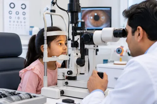
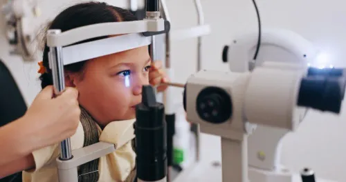
- Read an eye chart for visual acuity, then undergo slit-lamp examination of the cornea, iris, lens and other front-of-eye structures.
- Have intraocular pressure measured. One normal reading does not rule out glaucoma, and raised pressure alone does not prove nerve damage.
- Undergo gonioscopy, using a special contact lens to inspect the drainage angle. It distinguishes open-angle glaucoma from angle-closure glaucoma and helps identify secondary glaucoma caused by steroids, trauma, inflammation or lens changes.
- The clinician examines the optic nerve for cupping and other damage.
- Complete visual-field testing to detect missing peripheral vision.
- Have optical coherence tomography (OCT) map the retinal nerve-fibre layer and ganglion-cell layer.
- Receive corneal-thickness measurement when relevant, because thickness affects pressure interpretation.
This sequence matters in glaucoma care in mumbai: treatment depends on the angle, nerve, field and imaging together, not symptoms or pressure alone.
How do visual fields and OCT reveal damage that you cannot feel?
A visual-field test detects missing peripheral vision before you notice it. You sit facing a bowl-shaped instrument, keep looking at a central target, and press a button whenever a light appears, even when it is faint.
Reliability matters because the result can look worse or better than your actual field:
- Fixation losses suggest you looked away from the central target.
- Missed responses can reflect fatigue, poor understanding or a genuine field defect.
- False responses, given when no light appeared, can make the field look artificially normal.
Bring your current glasses, explain if the instructions are unclear, and rest between tests. A repeat field is useful when you are tired, learning the task, responding inconsistently or producing a result that does not match the optic-nerve examination. One unreliable test should not determine glaucoma vision treatment.
OCT uses light to map the retinal nerve-fibre layer around the optic nerve and the ganglion-cell layer in the macula. Comparing scans over time can reveal thinning that is progressing, even while central vision feels normal.
An OCT printout alone cannot confirm or exclude glaucoma. Your clinician must interpret image quality, eye pressure, drainage angle, optic-nerve appearance and visual-field results together.
How do doctors separate glaucoma from cataract, retinal disease and ordinary blur?
Spectacles can sharpen blur caused by an uncorrected refractive error, but they will not repair optic-nerve damage. Choosing adult glaucoma treatment therefore requires more than asking whether letters look clearer.
| Pattern | Clue that points elsewhere |
|---|---|
| Refractive error | Vision improves with the correct spectacles. |
| Cataract | Glare, reduced contrast and difficulty seeing in dim light are prominent; cataract treatment may improve these symptoms. |
| Diabetic retinopathy | Retinal changes, bleeding or swelling appear on examination. |
| Macular disease | Central vision becomes distorted, blurred or missing. |
| Dry eye | Blur fluctuates and often improves after blinking or lubricating drops. |
| Optic-nerve disease | Field defects or abnormal colour vision can occur. |
Improvement after spectacles or cataract treatment does not exclude a separate glaucoma problem. A person can have clearer central vision while retaining unnoticed peripheral loss from glaucoma.
Doctors assess glaucoma through the optic nerve and visual field, not symptoms alone. They also compare eye pressure, drainage angle and retinal imaging with the examination findings. A normal pressure reading does not rule it out, and raised pressure does not prove nerve damage.
A field defect or colour-vision change can point to optic-nerve disease, while retinal examination identifies diabetic or macular causes.
Which glaucoma treatments can protect vision that remains?
Glaucoma vision treatment protects the sight that remains; it cannot restore optic-nerve damage already caused by glaucoma. Your ophthalmologist sets an individual target pressure based on the damage, eye structure, age and risk of progression. Treatment is adjusted if the pressure meets its target but visual fields or the optic nerve continue to worsen.
| Option | Main advantage | Important limitation |
|---|---|---|
| Daily eye drops | Lower pressure without a procedure | Missed doses, incorrect instillation or stopping because vision feels unchanged can allow silent progression |
| Selective laser trabeculoplasty | Can reduce dependence on daily drops in suitable open-angle glaucoma | Its effect can diminish; medication or surgery may later be needed |
| Trabeculectomy or drainage device | Produces substantial pressure reduction | Requires close postoperative monitoring and carries risks including low pressure, infection, inflammation, scarring and further procedures |
| Less-invasive glaucoma procedures | Smaller surgical intervention | Usually offers a more limited pressure reduction |
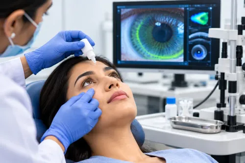
Tell your doctor about asthma, a slow pulse or heart-conduction problems before using a beta-blocker drop. Prostaglandin analogues can cause redness, eyelash growth, and darkening of the iris or skin around the eye.
- Wash your hands, place the prescribed drop correctly, and ask whether eyelid closure or tear-duct pressure is needed afterward.
- Ask what to do if the bottle tip touches your eye or you miss a dose.
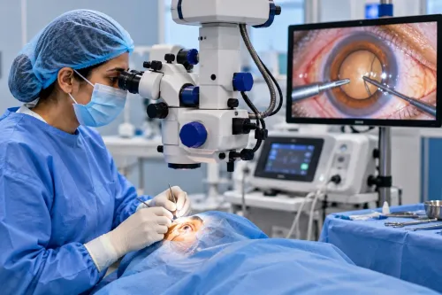
- Discuss laser or surgery when pressure stays above target, testing shows progression, or drops and laser are unsuitable.
Never change treatment because your vision feels unchanged; that absence of symptoms does not prove the disease is stable.
How should you choose glaucoma care in Mumbai for gradual change?
Choose a glaucoma specialist in Mumbai who will investigate the cause of gradual change, not treat every case as routine open-angle glaucoma. Ask whether the consultation includes gonioscopy, optic-nerve examination, visual-field testing, pressure measurement, and, when useful, OCT and corneal-thickness measurement.
Use this checklist:
- Disclose a close relative with glaucoma, even if you have no symptoms; peripheral loss can develop unnoticed.
- Provide every systemic medicine and mention steroid use, previous eye trauma, inflammation or lens problems.
- Ask how pressure will be compared with the optic nerve and visual field. One normal pressure reading does not rule out glaucoma.
- Request a written follow-up interval and the tests that will detect progression.
| Mechanism | What it means | Why it changes care |
|---|---|---|
| Open-angle | Drainage angle remains open | Pressure and nerve damage guide treatment |
| Angle-closure | Drainage angle is narrowed or blocked | Urgent risk and different treatment choices |
| Secondary | Another condition or exposure contributes | Treating the cause affects the plan |
For an adult in Lower Parel, Clarity Eye Clinic-Dr.Nikita Mashru can be considered for a structured assessment or treatment discussion. Relatives also need periodic comprehensive eye examinations because age, eye anatomy and medical factors change risk; family screening is not a one-time clearance.
Related service
GLAUCOMA TREATMENT Glaucoma Care by Dr. Nikita Mashru Glaucoma Care at Clarity Eye Clinic, Lodha Signet, Lower Parel, Mumbai, focuses on the early detection, diagnosis,... View service → |
Frequently asked questions
Can gradual vision changes indicate glaucoma?
Yes. Glaucoma can reduce peripheral vision gradually without pain, while blur, glare and poor dim-light vision can also indicate cataract or refractive error. Arrange an eye examination to identify the cause.
What happens during an adult glaucoma assessment?
The assessment can include eye pressure measurement, optic nerve examination, visual field testing, OCT imaging and checks for cataract, retinal disease and refractive error.
How do visual fields and OCT detect glaucoma damage?
A visual field test maps areas of reduced sensitivity, including side vision. OCT measures the thickness and structure of retinal nerve fibres and the optic nerve, revealing changes you may not feel.
Which treatments can protect vision affected by glaucoma?
Treatment can include prescription pressure-lowering eye drops, laser procedures or glaucoma surgery. The choice depends on eye pressure, optic nerve damage, disease progression and response to treatment.
How should you choose glaucoma care in Mumbai?
Choose a glaucoma specialist in Mumbai who offers complete testing, explains the diagnosis and treatment options, monitors progression over time, and provides access to laser or surgical care when needed.
Keywords